Understanding Chiari Malformation Causes and Effects
Chiari malformation is a neurological condition where brain tissue extends into the spinal canal, interfering with normal cerebrospinal fluid flow. This structural abnormality, often present at birth, can lead to a range of symptoms, including headaches, neck pain, balance problems, and difficulty swallowing. Though its severity and impact vary among individuals, understanding Chiari malformation is vital for timely diagnosis and management. With advancements in medical research and treatment, patients can access various therapeutic options to improve their quality of life and alleviate symptoms associated with this condition.
Causes of Chiari Malformation

Chiari malformation can arise from a variety of factors, which are typically categorized as congenital or acquired causes. Below is an exploration of these primary causes.
Congenital Causes
Congenital causes of Chiari malformation typically originate during fetal development, leading to structural abnormalities in the skull or brain. Below are the primary factors contributing to congenital causes.
Genetic Factors
Genetic predisposition may play a role in some cases of Chiari malformation. Mutations or inherited disorders can result in abnormal bone development, especially affecting the structure of the skull. This can lead to insufficient space for the brain, causing the cerebellum to push downward into the spinal canal. Research continues to explore how hereditary factors influence the development of Chiari malformation.
Developmental Issues
During fetal growth, improper development of the brain and surrounding structures can lead to congenital Chiari malformation. This often involves the underdevelopment of the bones at the skull’s base, causing crowding in the brainstem and cerebellum regions. Such developmental anomalies can result from genetic, environmental, or unknown factors affecting fetal growth.
Acquired Causes
Acquired Chiari malformation develops later in life due to certain conditions or factors that alter the structure of the skull or brain.
Head or Neck Trauma
Severe trauma to the head or neck can lead to structural changes in the skull or spinal column, causing the brain to be pushed downward. Such injuries may disrupt the flow of cerebrospinal fluid, exacerbating symptoms of Chiari malformation over time. Prompt medical evaluation after trauma is essential, as early intervention can mitigate long-term complications associated with this condition and improve the patient’s prognosis.
Excessive Spinal Fluid Drainage
Certain medical conditions, such as hydrocephalus or meningitis, require treatment methods involving the drainage of cerebrospinal fluid. Excessive or rapid removal of this fluid can lead to a vacuum effect, pulling the brain downward into the spinal canal. This can result in the development of symptomatic Chiari malformation, even in individuals with no prior abnormalities. Monitoring fluid drainage carefully during treatment is crucial to prevent this complication.
Types of Chiari Malformation
Chiari malformation is categorized into several types, each defined by the severity of the condition and the anatomical structures involved.
Type I Chiari Malformation
Type I is the most common form, often diagnosed in late childhood or adulthood. It occurs when the lower part of the cerebellum, known as the cerebellar tonsils, extends into the spinal canal. Symptoms may include headaches, neck pain, balance issues, and neurological deficits. Many individuals remain asymptomatic, and the condition is frequently discovered incidentally during imaging performed for unrelated concerns. Early diagnosis allows for better symptom management and assessment of potential complications.
Type II Chiari Malformation
More severe than Type I, Type II is typically present at birth and often occurs with spina bifida. This form involves a greater portion of the cerebellum and brainstem being pushed into the spinal canal. Symptoms can include breathing difficulties, feeding problems, and structural abnormalities of the spine and skull. Because of its association with other neural tube defects, treatment often necessitates a multidisciplinary approach involving neurosurgeons and specialists. Early intervention is crucial to improve quality of life for affected individuals and to address coexisting conditions.
Type III Chiari Malformation
The rarest and most serious type, Type III is characterized by the cerebellum and brainstem protruding into an abnormal opening at the back of the skull. This leads to significant neurological impairments, including seizures, delayed development, and muscle weakness. Survival rates are lower due to the complications it presents, and surgery is often required to repair the defect. Cases are typically diagnosed through prenatal imaging or shortly after birth. Managing this severe condition requires a combination of medical care and supportive therapies to address developmental challenges.
Symptoms of Chiari Malformation
Chiari malformation symptoms can vary widely depending on the type and severity of the condition. Common symptoms include:
- Headaches, often worsening with coughing or straining
- Neck pain or discomfort
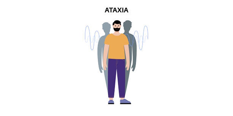
- Balance and coordination issues
- Weakness or numbness in the limbs
- Difficulty swallowing or speaking
- Breathing problems in severe cases
- Tinnitus or hearing disturbances
- Spinal abnormalities, such as scoliosis
Diagnosis and Imaging Techniques

Diagnosis of Chiari malformation typically involves a combination of medical history, physical examination, and advanced imaging technology to confirm the condition.
Magnetic Resonance Imaging (MRI)
MRI is the gold standard for diagnosing Chiari malformation. This technique provides detailed images of the brain and spinal cord, allowing doctors to observe the position of brain structures, such as the cerebellum, and detect abnormalities. An MRI can also help assess related conditions like syringomyelia or hydrocephalus. It is a non-invasive procedure and is particularly valuable in guiding treatment decisions based on the severity of the malformation.
Computed Tomography (CT)
CT scans, though less detailed than MRIs, can also play a role in diagnosing Chiari malformation. They use X-rays to create cross-sectional images of the brain and spine, focusing on bone structures. CT scans are especially useful for identifying bony abnormalities or malformations at the base of the skull and spine. Additionally, in emergency situations, CT imaging is quicker and more accessible, making it effective for preliminary evaluations when Chiari malformation is suspected.
Neurological Examination
A comprehensive neurological examination is crucial alongside imaging tests. This includes assessments of reflexes, coordination, balance, and motor skills. These tests help identify the functional impact of Chiari malformation, offering insights into the severity of symptoms. Through neurological evaluations, physicians can detect subtle deficits in movement, sensation, or cranial nerve function, guiding further diagnostic steps and the development of a personalized treatment plan for each patient.
Conclusion
Chiari malformation is a complex condition requiring careful diagnosis and management. While imaging tests like MRIs and CT scans provide crucial insights into anatomical abnormalities, neurological examinations focus on identifying functional impairments. Together, these tools enable physicians to make accurate diagnoses and tailor treatment plans to each patient’s specific needs. Early detection and intervention are vital to managing symptoms and preventing complications.


